3D Printing: To Make a Tissue Out of Technology
By Rajaneesh K Gopinath, Ph.D.
Tissue engineering is a subfield of regenerative medicine that confronts numerous technical challenges. 3D printing could well be the leap in technological innovation that reignites its ambitions.
Introduction
The dawn of tissue engineering as a budding discipline occurred during the late 1970s, when after a series of unsuccessful attempts, Dr. W. T. Green rightly envisioned the need for improved biomaterials as scaffolds for seeding cells (1). However, the landmark article published by pioneers, Dr. Robert Langer and Dr. Joseph Vacanti is often referred to have marked the inception of the field (2). Their article summarized in detail the different mammalian tissues that were attempted to be engineered using contemporary methods at the time.
Biodegradable three-dimensional (3D) scaffolds
 By 1984, Charles W. Hull had invented the first 3D printing (3DP) technology called Stereolithography, a process by which thin layers could be printed successively to make a solid object. Within a few years, tissue engineering had incorporated the 3DP technique. Any engineered tissue has to provide structural and mechanical support, nutrients to the cells and get activated once implanted (3). So, the success of tissue engineering largely rests on building optimal scaffolds that resemble the native extracellular matrix (ECM). Therefore, the major goal is to design an ideal porous 3D scaffold that accommodates cell migration and growth. Although numerous techniques were perfected in developing 3D scaffolds, it was still challenging to attain consistent pore size and control of scaffold architecture (4). It was this limitation that led to the use of 3DP, which facilitated the regulation of pore size and structure. In comparison to existing methods, 3DP offered various advantages. It was possible to incorporate pharmaceutical agents into the scaffolds and create complex drug delivery systems (5). Besides, it offered the comfort of multi-“color” printing, with which multiple cells could be arranged simultaneously (6).
Hydrogel Scaffold System
Hydrogel has proved to be a very attractive 3DP scaffold since they are biocompatible, could be easily controlled and more importantly, mimic the ECM environment. They are hydrophilic polymer chain systems that have high-water content and their nutrient and waste exchange systems have similar diffusion properties (7). The major component of Hydrogel is gelatin, while glutaraldehyde is used as a crosslinking agent to increase stability. To overcome the toxicity of crosslinkers, controlled gelation agents such as alginate is sometimes used in combination with calcium chloride. Several techniques such as molding and soft lithography among others have been used to fabricate hydrogel constructs. Not long ago, a new fabrication method called femtosecond laser-induced two-photon polymerization (2PP) has emerged as a nanoscale 3DP technique that could create 3D hydrogels with more precision in terms of its size and structure (8).
3D-Bioprinting
The past decade witnessed the extensive evolution of 3DP techniques to a degree that a new subfield called 3D bioprinting has emerged. Clemson University received the first patent in this field in 2006 for the invention of ink-jet printing of viable cells. Around the same time, a technique called “Organ printing” was proposed by Vladimir Mironov and coworkers. This technique comprises three sequential steps; 1) Making an organ blueprint, 2) organ printing and 3) organ conditioning and maturation (9). By placing microtissues in the form of spheroids as building blocks, tissue assembly could be promoted by self-fusion (10). Mironov later went on to become the chief scientific officer of 3D Bioprinting Solutions. This start-up company designed the first Russian multifunctional 3D Bioprinter, Fabion that printed the first-ever functional organ, the mouse thyroid gland. Yet, Organovo, a San Diego based bioprinting company was the one to set precedent in commercializing 3D bioprinting technology. In 2014, they launched the ExVive™ 3D bioprinted human liver tissues that could be used for drug testing and metabolism studies (11). What’s more, they have now developed a system where bioink could be obtained from donor organs for printing. The ultimate challenge would be to print whole organs that could be transplanted into patients and resolve the issue of donor unavailability in the future.
Innovations and Future Challenges
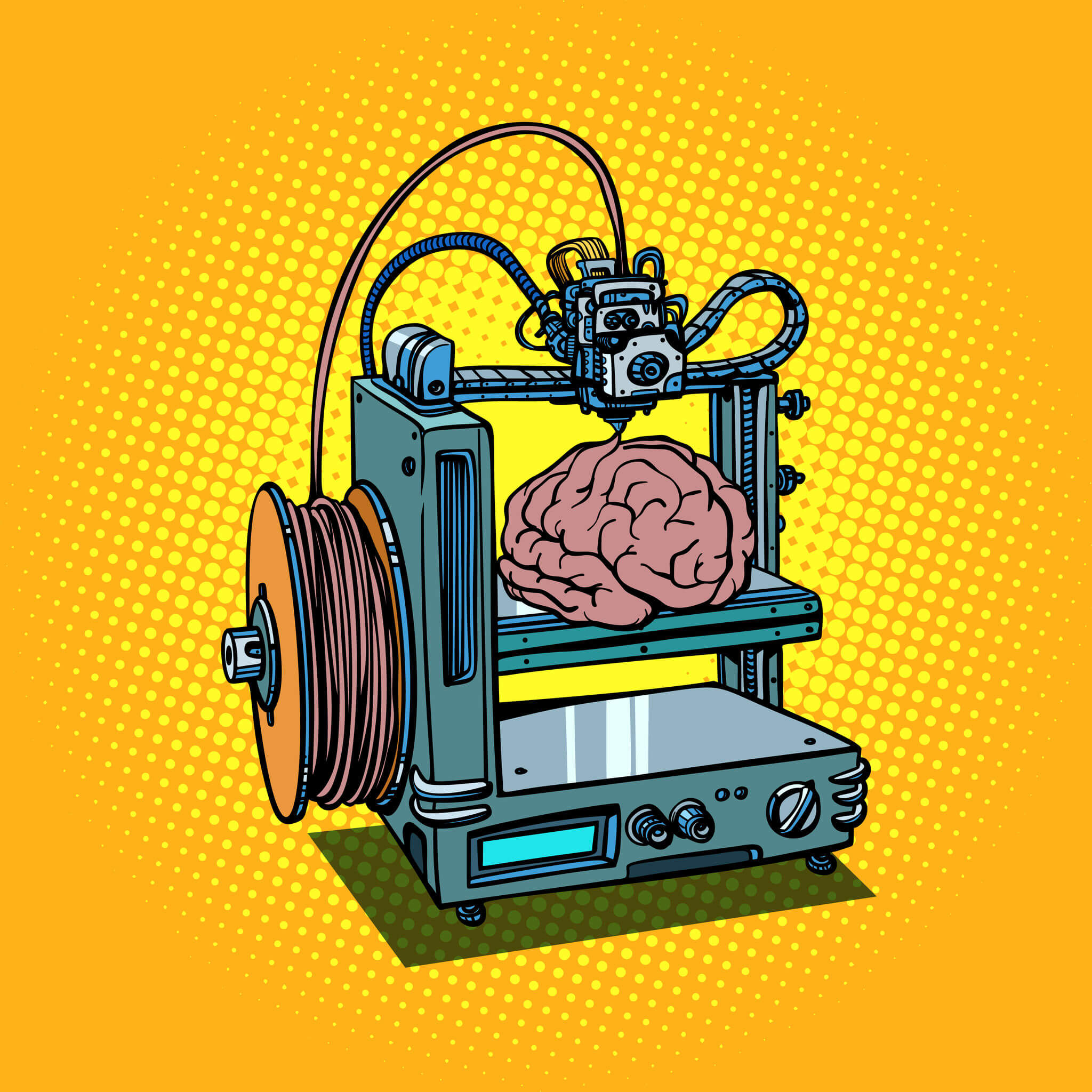
Recently, a novel 3DP technique called 3D cryogenic printing is developed by scientists in the UK using hydrogels (12). This technique produces stable 3D structures using a composite hydrogel ink that could be cooled rapidly using solid carbon dioxide once it is printed. This innovation creates structures that closely resemble soft body tissues like the brain thereby rendering promise to studies of complex brain injuries. The biggest challenge now in the field is to translate the success of ex vivo 3D printed structures to function in vivo. The ultimate challenge would be to print whole organs that could be transplanted into patients and resolve the issue of donor unavailability in the future. Besides, 3D printed tissues are proving to be a good alternative that replaces animal models in testing chemical compounds and pharmaceutical drugs.
References
- https://www.ncbi.nlm.nih.gov/pubmed/16989721
- https://www.ncbi.nlm.nih.gov/pubmed/8493529
- https://www.ncbi.nlm.nih.gov/pubmed/15675684
- https://www.sciencedirect.com/science/article/pii/S2095809916300716
- https://www.sciencedirect.com/science/article/pii/0168365995001735
- https://www.ncbi.nlm.nih.gov/pubmed/22692615
- https://www.ncbi.nlm.nih.gov/pubmed/19472329
- http://pubs.rsc.org/-/content/articlehtml/2018/tb/c8tb00301g
- https://www.ncbi.nlm.nih.gov/pubmed/12679063
- https://www.ncbi.nlm.nih.gov/pmc/articles/PMC3773699/
- https://www.ncbi.nlm.nih.gov/pmc/articles/PMC5491216/
- https://www.ncbi.nlm.nih.gov/pmc/articles/PMC5701203/
©www.geneonline.com All rights reserved. Collaborate with us: service@geneonlineasia.com

Author
LATEST

EVENT
2024-04-20
16th SABPA OC/LA Annual Biomedical Forum
The Beckman Center, 100 Academy, Irvine, CA, 92617
2024-04-27
2024 Biomedical Final Pitch Competition
Room DA1620, Dana Building, Dana-Farber Cancer Institute, 99 Jimmy Fund Way, Boston, MA 02115